If you stare at an object with steady eyes, you can see it clearly. Even without moving our eyes in any other direction, we perceive a great deal of things around it too. This whole perceptual area is called the visual field..
We distinguish between a monocular field of view (which we see with only one eye) and a binocular field of view. In the middle, centrally, the visual field of the two eyes overlaps. During the field of vision examination, the individual’s field of vision is mapped.


The field of vision
The visual field is an indicator of glaucomatous damage. Visual field defects may initially develop in the outer parts (periphery), but may also occur near the centre of the visual field. The patient’s ability to recognise this is almost impossible, especially if these processes occur slowly.
Due to the coincident visual field defects in the two eyes with central visual field loss, there is a risk that the visual field defect in one eye is compensated by the other eye, and the patient may not notice the damage.
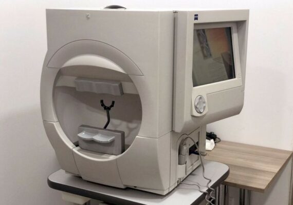
Glaucomatous damage continues to progress unnoticed, until the same visual field area is lost in both eyes. To detect visual field loss in time and to monitor its progress, a very important device is the Visual Field Perimetry Testing Equipment.
Computer examination
A patient with glaucoma requires eye care:
Glaucoma is a chronic, slow-moving disease in which the number of optic nerve fibres is constantly reduced. The destruction of nerve fibres causes a narrowing of the visual field and permanent damage to vision. The aim of regular eye check-ups for glaucoma patients is to help preserve eye function. Only timely and appropriate therapy can preserve vision. Internal medical risk factors should also be monitored throughout the course of the disease. Internal medical risk factors should also be monitored throughout the course of the disease.
How is the visual field test performed?
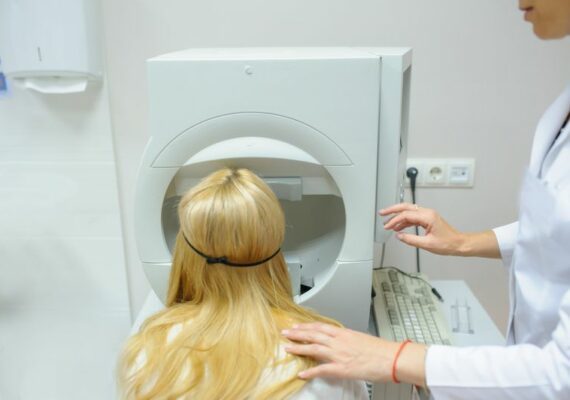
During a painless visual field test, we map the individual’s field of vision in each eye separately. Using a visual field testing (perimetry) machine, the patient sits in front of a large hemisphere and the eye being tested looks exactly at the centre of the hemisphere. The head is fixed by the chin and forehead support. The gaze is fixed on the mark in the centre of the hemisphere. Inside the hemisphere, small points of light appear at different places, and the patient indicates their appearance by pressing a switch.
On the earlier devices, the doctor moved the small spot of light mechanically, but on modern perimeters the computer controls the appearance of the light spots and registers the patient’s feedback. The perimeters can be used to detect visual field loss and its extent.

